Important Diseases Diagnosed and Treated in the Department of Cardiology: Heart attack (acute myocardial infarction), coronary insufficiency (chronic ischemic heart disease), heart failure, heart rhythm and conduction disorders, heart valve diseases, peripheral vascular diseases, aortic vessel diseases, hypertension, hypercholesterolemia, congenital heart diseases observed in adulthood. Diagnosis and Treatment Methods used in Our Hospital Electrocardiography (ECG) Effort (Treadmill) Test Echocardiography Transoesophageal Echocardiography ECG Holter Blood Pressure Holter Tilt-Table Test Coronary Angiography Peacemaker Implantation Additionally, imaging of kidney vessels, neck veins and leg veins and if necessary, intervening in the occluded vessels by means of balloon and stents, are among the diagnostic and treatment methods applied in the angiography unit. The renal denervation method is used for patients who are monitored for hypertension and whose blood pressure cannot be kept under control despite drug treatment. TAVI (Transcatheter Aortic Valve Implantation): In patients with aorta valve stenosis, aorta valve replacement can be performed by entering the heart through the groin vessel by TAVI, which is a non-surgical valve replacement method. MitraClip: This procedure is intended to treat the insufficiency of the mitral valve between the left atrium and the left ventricle in the hearts of patients with high surgical risk. In the event of a blood leakage from the mitral valve, a latch is inserted into the heart by entering the heart through a process similar to angiography with the help of a catheter. After 2 – 2.5 hours, patients can return to their daily lives. EPS/Ablation: An electrophysiological study (EPS) is performed on patients diagnosed with fainting, palpitations and arrhythmia. In patients with arrhythmia, treatment is called ablation, which is a non-surgical procedure and is performed by entering the groin vein in order to reach the heart. With the latest technology devices available in the angiography unit, all kinds of rhythm disorders can be diagnosed and treated. ILR (Implantable Loop Recorder): In the patient group who have fainting or palpitation attacks and who cannot be diagnosed with Holter and similar tests, long-term cardiac rhythm recordings can be made with an ILR device placed under the skin with an incision of approximately 1-2 cm in the epigastric region, and many cases of arrhythmia can be diagnosed and treated. ASD / VSD / PFO Closure: ASD / VSD / PFO are congenital holes between the compartments of the heart. The heart holes of patients who are suitable for the procedure can be closed with the help of a plug-like apparatus without the need for surgery. Virtual Angiography: The SIEMENS Somatom Definition Flash computer tomography device in the radiology unit provides information about the cardiovascular system of people suspected to have cardiovascular disease without applying any invasive procedures. SUB DEPARTMENTS OF THE CARDIOLOGY DEPARTMENT Effort Test Laboratory: Effort and exercise testsare usually done on treadmills, where cardiac workload is increased gradually. In the meantime, the EKG is monitored continuously and blood pressure is measure periodically. The Effort test is performed in order to diagnose atherosclerosis, evaluate the drug effects and effort capacity of diagnosed diseases and to investigate rhythm and conduction impairments, which can only be determined with exercise. Holter Laboratory: Arhythm Holter records the heart rhythm for 24 hours with a small device that is connected to electrodes attached on the chest wall. It is done in order to determine cardiac rhythm and conduction impairments, evaluate the treatment success of arrhythmia, examine the relation between fainting and electrical activities of the heart and finally, to examine silent coronary artery disease. The Holter device measures blood pressure for 24 hours automatically at certain intervals. It can be used for hypertension diagnosis and for evaluating the efficiency of the treatment. Telemetry Unit: It is a method for the follow-up of patients with rhythm or conduction disorders who are not bedridden but are currently being treated in the hospital. The rhythm of the patient, who carries a small recording device connected to the electrodes, can be continuously monitored at the Telemetry Unit and recorded if necessary. Echocardiography Laboratory: Echocardiography is the process of examining the structures and functions of the cardiac valves, heart muscle and dimensions of the cardiac cavities with the help of sound waves. The most common way of doing this is through transthoracic echocardiography (TTE). In this method, an ultrasonic probe is used on the chest cage from the outside for examination.Transoesophageal echocardiography (TEE) is done by placing an ultrasonic probe into the oesophagus. Because of the close proximity of the heart and oesophagus, this method can clearly visualize the cardiac cavities, aorta and cardiac valve structures. TEE is the method of choice for evaluating clots, aortic ruptures in cardiac chambers, infections of cardiac valves, heart perforations and prosthetic valves. Stress echocardiographyis an echocardiography procedure performed after the heart workload is increased with effort or certain medication. It is designed to investigate coronary artery disease and to investigate the cardiac muscle more precisely after a heart attack. Coronary Intensive Care Unit: The Department of Cardiology has a 24-hour Coronary Intensive Care Unit equipped with the latest technologies. Severe heart problems such as heart attack, acute heart failure, and cardiogenic shock are closely monitored and treated in this unit. Temporary cardiac pacemaker, catheterization of the pulmonary artery, intra-aortic balloon and mechanical ventilation are some of the medical procedures performed in the coronary intensive care unit. Nuclear Cardiology Unit: Radionuclide Ventriculography is the process of measuring the blood pumping function of the heart by means of a very small dose of radioactive material delivered from the vein. Even a slight reduction in the contraction function of the heart can be detected by this sensitive method. Coronary Artery Disease can be diagnosed with more than 95% accuracy viaPositron Emission Tomography (PET)and it can be evaluated precisely whether the patients who have had a heart attack and regional damage to the heart muscle can benefit from Balloon Angioplasty stent or an Aortocoronary Bypass operation. Heart Catheterization and Coronary Angiography Laboratory: In the Angiography Laboratory, detailed information about heart pumping function, cardiac valves and coronary vessels is obtained. Important decisions related to coronary bypass operations, balloon angioplasty and cardiac valve operations are taken on the basis of this information. Coronary Angioplasty and Stent Application: Coronary angiography is usually treated with Percutaneous transluminal coronary angioplasty (PTCA) or stent if the strictures detected in the coronary vessels are at the state where they are preventing blood flow. Similar to Coronary Angiography, the PTCA operation can be performed by entering through the artery in the leg or arm. Although the success rate of PTCA treatment is high, the same vascular recurrence may be observed in the first 6 months. For this reason, metallic material called a "Stent" is applied by mounting it on a balloon catheter and it is known to decrease the narrowing rate and is therefore used frequently today.
The Cardiology Department of Near East University Hospital is working to protect heart health, as well as to diagnose and treat existing heart diseases based on world standards.
It is used to study the function of the heart muscle and neural transmission system. The resulting electrical changes are recorded by means of conductive leads (electrodes) placed in various parts of the body. ECG helps in the diagnosis of many cardiological diseases such as cardiovascular diseases, heart muscle diseases and rhythm disorders.
The Effort test is a frequently used test to investigate the presence of cardiovascular diseases, to determine the efficacy of treatment in known cardiovascular diseases, to diagnose rhythm disturbances associated with exercise and to investigate the exercise capacity of a person in some cases of cardiovascular diseases. The Effort test is particularly beneficial in early diagnosis and diagnosis of heart diseases.
One of the most widely used echocardiography devices in the heart centre, the Vivid E9 echocardiography device, enables 2 and 3-dimensional heart imaging, doppler methods, heart valves and detailed blood flow measurements can be made.
Transoesophageal echocardiography, which is an endoscopic examination, is used to reach the posterior region of the heart through a thin tube (probe) which is lowered from the mouth to the oesophagus, so that the cardiac cavities and heart valves can be examined in detail. The TEE procedure is performed with the Vivid E9 echocardiography device and special apparatus, and 2-D and 3-D images of the heart valves and heart cavities can be obtained when necessary.
With the ECG Holter, a device similar to a mobile phone and attached to a belt, the heart electrocardiogram can be recorded during a planned period of time while the person maintains a normal daily life. With this device, all heart rhythm disturbances such as palpitations, chest pains, and sensations of fainting can be detected, which are not seen during the examination but can happen for a short duration during the day.
These devices measure blood pressure for a 24-hour period with 15 – 20minute intervals, and 24 hours of blood pressure monitoring can beconducted, especially for people who areat risk of hypertension or those who cannot be controlled with medication.
It is a test used for the diagnosis of syncope. The Tilt Table Test is performed in an outpatient clinic on a tiltable table. Blood pressure and pulse are monitored at regular intervals and clinical conditions that may cause fainting are investigated.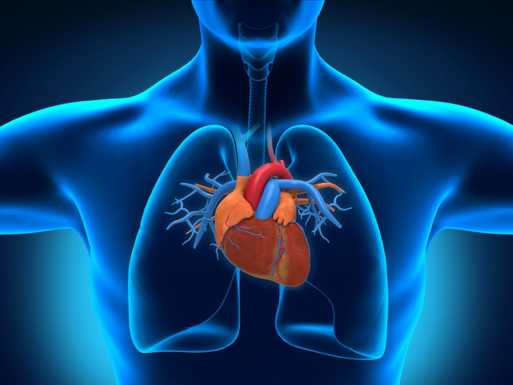
Interventional Procedures - Angiography Unit
GE Innova 3100, one of the newest devices, is used for coronary angiography to evaluate cardiac vessels, and if there is severe stenosis in the vessels, balloon or stent placement is performed.
In addition, various pacemakers such as KPM, ICD, CRT are inserted with the same devices in the angiography unit. Cardiac pacemaker controls of patients who have had a Medtronik brand battery device implanted at the Heart Centre are performed by cardiologists who are experts in this field.
Cardiac Check-up Unit: Acheck-up is a health check that is performed in order to diagnose diseases before complaints appear. During the check-up process, the risk factors of heart diseases are examined, a physical examination is performed, an effort test is applied, and echocardiography and blood test results are evaluated. If the results point to a possible heart disease, further examinations are required for final diagnosis. If the examinations are all normal, recommendations are given to the patient in order to reduce the risk factors and preserve heart health.2. Floor Eastern Block Department of Polyclinics Monday - Friday 8:00 - 17:00 Saturday Closed Sunday Closed +90 (392) 444 0 535
Policlinic Area
Opening Hours
Location
Department Doctors