The technological equipment available in the Radiology Department enables all diagnosis and interventional radiological services. The digital images obtained from our advanced technological devices are recorded and archived in the Picture Archiving and Communication System (PACS). This helps to obtain more detailed and higher quality images in a faster manner in comparison to other methods of imaging, and these images are evaluated by specialist doctors in the PACS work stations. All the services expected from a fully - equipped radiology unit are available at our clinic. These are:
- Digital radiography (DR) system
- Digital Fluoroscope
- Digital Mammography
- Ultrasonography, Doppler and 4D Ultrasonography
- Multislice Computed Tomography
- Magnetic Resonance Imaging (1.5 Tesla, with 48 channels)
- Digital Angiography (DSA)
- Interventional Radiology
- Image Storing and Transmission System (PACS)
Digital Radiographic Examinations
All conventional imaging can be obtained with the DR systems and all routine and specific direct imaging examinations (scoliosis, distance measurement and cephalometry) can be conducted. Additionally, if needed, patients are put through a Mobile X-Ray Devices, which is a radiography service that can be conducted when the patient is lying down.
Devices used:
- GE Definium (TM) 6000 Digital Radiography Systems
- Siemens Mira Max Digital Mobile Radiography Systems
Digital Fluoroscope Examinations
All gastrointestinal system imaging (oesophagus, stomach, duodenum, small intestine and double contrast colon imaging), urological radiological tests (IVP, cystography), hysterosalpingography (HSG), and fistulography examinations can be conducted.
Device used: Siemens Luminos Fusion Fluoroscopy Device.
Digital Mammography Examinations
Digital mammography, which is a new technology used for breast imaging, has become the main breast cancer detection and diagnosis method. Different from the classical mammography, it obtains high quality images with lower doses of radiation and sensitive electronic detectors in a digital environment. Then, these images can be examined on work stations with high resolution monitors. Image processing procedures such as contrast settings can be adjusted regardless of the enlargement, measurement and dose of X-rays used.
Advantages of Digital Mammography
- It is quick. The images can be obtained immediately after the imaging and the validity of these images can be detected.
- The dose of radiation is a lot less than the classic mammography.
- Its contrast resolution is higher than the normal mammography. Microcalcification and small lesions can be differentiated easier. This helps to examine the dense and fibrocystic breast tissue.
- The images can be digitally archived and transferred.
Device used: Siemens Mammomat Inspiration.
Ultrasonography and Doppler Ultrasonography Examinations
The high-resolution imaging quality of the ultrasonography devices helps diagnosis. We provide services with our latest technology ultrasonography devices in specially prepared rooms. All ultrasonographic imaging and interventional procedures accompanied by ultrasonography are applied at our department. In particular, pregnancy ultrasound and colour 4 D imaging can be conducted.
Devices used:
- GE LOGIQ S 8 Ultrasonography and Doppler Ultrasonography Device
- Siemens Acuson S2000 Ultrasonography and Doppler Ultrasonography
- GE Voluson 730 Pro Ultrasonography and Doppler Ultrasonography Device
- Siemens Acuson NX3 Ultrasonography and Doppler Ultrasonography
Multi-Slice Computed Tomography Examinations
“Multiple Detector CT” with 256 dimensions enables faster and higher resolution images to be taken. The examinations that can be conducted with these devices, which are equipped with special imaging programs for each region, include:
- Conventional examinations for full body regions
- 3D imaging
- CT-Angiography (cerebral, carotid, aorta, peripheral angiogram) examinations
- Coronary CT-Angiography and Calcium Scoring
- Full body screening (trauma panel)
- Research and procedures for patients who present to the emergency service with chest pain (aorta dissection, pulmonary embolism, pneumonia etc.) are successfully conducted.
Device used: Siemens Definium Flash 256 Section IT Device.
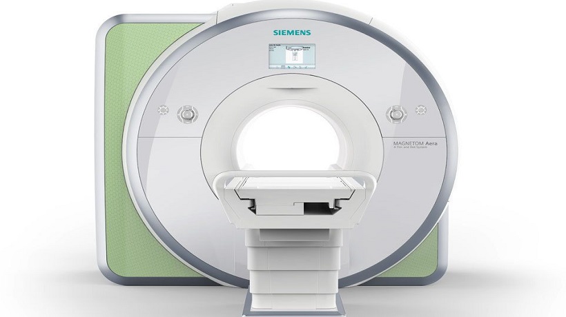
Magnetic Resonance Imaging (MRI)
Near East University Hospital
- Head focussed imaging such as: brain, eye, internal ear and ear structures, hypophysis, jaw joint, brain vessels and artery systems
- Contrasted (DSC and DCE) and non-contrasted (ASL) perfusion MRI
- Diffusion and diffusion tensor imaging
- Functional brain MR examinations
- MR-Spectroscopy examinations
- Brain spinal fluid flow research
- Neck structure, throat, saliva glands, tongue and surrounding structure examinations
- Lung, heart and cardiovascular examination
- Morphologic and functional cardiac imaging, Coronary MR-Angiography and Cardiac perfusion examination
- Contrast and non-contrast MR angiography for full body
- Neck, back, waist spine and MR myelography examinations
- Morphological and kinematical examination of shoulder, arm, elbow, wrist, hand, hip, femur, knee, leg, ankle and foot joints
- Organs within the stomach and pelvic region examinations
- Dynamic tissue (lung, breast, kidney, tumour) MRI
- External cranium and abdominal diffusion applications
- MRCP (MR imagine technique used on bile duct and bile duct) examination
- MR-Pyelography (MR-Urography) examination
Device used: Siemens Magnetom Aera 48-channel MRI Device.
Digital Angiography
The monoplane Digital Angiography (DSA) device updated in 2015 at the Near East University Hospital enables venous and neurovascular interventional radiology applications at the latest standards. During 3D rotational evaluation of neuro-interventional methods using contrast material, the ‘’bolus-chase’’ method is used to evaluate the peripheral veins and with just one contrast material, the veins and arteries in both legs can be seen along the whole trace.
Non-invasive interventions:
- All forms of cerebrovascular angiography examination
- Inserting a stent into the narrow carotid to widen it
- Opening up blood clots and blocked arteries as a stroke intervention
- Aneurism treatment with endovascular method.
Vascular procedures:
- All forms of arteriovenous angiographic examination
- Reducing narrowness with intervention into the iliac and peripheral arteries and unblocking veins.
- Inserting a port and permanent dialysis catheter through the central vein.
- Renal arteriovenous fistula embolization.
Percutaneous method:
- Drainage catheter is placed and if needed, a stent is inserted in cases where intervention in the bile ducts is required
- Inserting a nephrostomy catheter with renal intervention.
Device used: GE 3100 Innova Angiography Device
Interventional Radiology
Interventional Radiology procedures are generally separated into three groups: vascular, non-vascular and neurovascular procedures.
Non vascular interventions include percutaneous (inserted from the skin) interventions. These interventions are conducted with ultrasonography or Computed Tomography. The most commonly used procedures are percutaneous drainage procedures performed with thin or thick needle mass biopsies. Additionally, nephrostomy procedures to relieve kidney canals that have widened, relieving bile ducts that have a tumour or narrowness and stent applications are conducted. Another procedure conducted with the percutaneous method is percutaneous laser application used to treating varicose veins without an operation.
Vascular interventions are conducted with the angiography device to open up narrow veins and blockages. Additionally, angioplasty and stent treatments are applied to the vascular structures of kidneys or bowels which have narrowness or sudden blockages. The most common venous application involves the insertion of a permanent tunnelled catheter into the central port of dialysis patients. Consequently, the dialysis patients can comfortably receive dialysis and cancer patients can comfortably have chemotherapy.
The neurovascular interventions conducted at our hospital include: neurovascular system angiography, inserting stent into patients with carotid narrowness, treatment of stroke patients who have been diagnosed at an early stage and treatment of patients who have brain embolisms due to aneurism.
Devices used:
- GE 3100 Innova Angiography Device
- GE Vivid E9 Echo Device
- GE Vivid S5 Echo Device
Picture Archiving and Communication System (PACS)
PACS (Picture Archiving and Communication System) is a web-based program which collates the images obtained from different imaging devices and then stores, prints if needs and files the images. As a result, images of the same patient taken with different devices can be stored and easily retrieved, old and new images can be compared, the images taken from external centres can be uploaded onto the system and these images can be shared between other dispensaries and health organisations connected to NEU on the web.
Additionally, PACS includes many FDA approved images within the system established at NEU Hospital and can be easily used in the cloud system. The main procedures conducted using this system includes:
- Basic applications used in daily examinations
- 4D program where dynamic images of drugs can be taken,
- Fusion program, which collates the images from different devices (CT, MRI, PET/CT) and stacks them on top of each other.
- 3D comparison program, which helps to compared both 2D and 3D images.
- Dental program, which helps evaluate dental images
- Virtual endoscopy program
- RECIST and PERCIST programs used for tumour monitoring and radiological and PET scan methods
- It is particularly used for forming 3D images, virtual operation plans or 3D modelling.
Device used: FUJİ PACS system
| 1. Floor | Western Block |
| Monday - Friday | 8:00 - 17:00 |
| Saturday | Closed |
| Sunday | Closed |